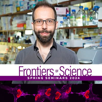
Frontiers of Science: Prof. Ricardo Henriques
When
Event Details
April 4th at 12:00
On-site event
Presidentti auditorium, BioCity
Spring 2024 program
Prof. Ricardo Henriques, Instituto Gulbenkian de Ciência, Portugal
Open Super-Resolution and AI technologies in the quest for nanoscale live-cell imaging
Host: Guillaume Jaquemet (guillaume.jaquemet@abo.fi)
Coffee and sandwich at 11:45, first come first served!
Six PhD researchers and early-career postdocs are welcome to have a lunch in restaurant Mauno and discuss with Prof. Henriques after the seminar. This is a great possibility to learn hosting skills in friendly environment and create connections for future. Everyone is welcome to join, BioCity Turku will offer the lunch.
If you got interested, please send an email to biocityturku@bioscience.fi
The combination of computational analysis and microscopy has transformed biological research, providing a powerful tool for quantitatively examining cellular processes. Advanced microscopy techniques, particularly those that rely on single-molecule localization for super-resolution imaging, require sophisticated analytical algorithms to interpret the vast amounts of data they generate. Our team, in collaboration with colleagues, has pioneered open-source frameworks that merge AI-based computational approaches and optical methodologies, specifically designed to enhance live-cell and super-resolution microscopy. These frameworks enable the high-performance acquisition of high-resolution quantitative data from biological specimens. In this presentation, I will showcase some of these advancements, including our platforms ZeroCostDL4Mic and DL4MicEverywhere, which make it easy for researchers to apply Deep Learning (DL) to the analysis of biological microscopy images, regardless of their coding skills. These platforms enable researchers to use cutting-edge DL techniques for tasks such as segmentation, object detection, denoising, and super-resolution reconstruction. We will illustrate the practical application of these open technologies through their use in investigating a range of biological phenomena, including the workings of eukaryotic and prokaryotic cells, as well as the analysis of single-molecule localization microscopy data. Through these demonstrations, we will explore how these technological advancements are not only pushing the boundaries of biological imaging but also empowering researchers to uncover new insights into the fundamental mechanisms of life.
Selected publications
Laine RF, Heil HS, Coelho S, Nixon-Abell J, Jimenez A, Wiesner T, Martínez D, Galgani T, Régnier L, Stubb A, Follain G, Webster S, Goyette J, Dauphin A, Salles A, Culley S, Jacquemet G, Hajj B, Leterrier C, Henriques R. 2023. High-fidelity 3D live-cell nanoscopy through data-driven enhanced super-resolution radial fluctuation. Nat Methods. 2023 Dec;20(12):1949-1956. doi: 10.1038/s41592-023-02057-w
Spahn C, Gómez-de-Mariscal E, Laine RF, Pereira PM, von Chamier L, Conduit M, Pinho MG, Jacquemet G, Holden S, Heilemann M, Henriques R. 2022. DeepBacs for multi-task bacterial image analysis using open-source deep learning approaches. Commun Biol. 2022 Jul 9;5(1):688. doi: 10.1038/s42003-022-03634-z
von Chamier L, Laine RF, Jukkala J, Spahn C, Krentzel D, Nehme E, Lerche M, Hernández-Pérez S, Mattila PK, Karinou E, Holden S, Solak AC, Krull A, Buchholz TO, Jones ML, Royer LA, Leterrier C, Shechtman Y, Jug F, Heilemann M, Jacquemet G, Henriques R. 2021. Democratising deep learning for microscopy with ZeroCostDL4Mic. Nat Commun. 2021 Apr 15;12(1):2276. doi: 10.1038/s41467-021-22518-0
Spark A, Kitching A, Esteban-Ferrer D, Handa A, Carr AR, Needham LM, Ponjavic A, Santos AM, McColl J, Leterrier C, Davis SJ, Henriques R, Lee SF. 2020. vLUME: 3D virtual reality for single-molecule localization microscopy. Nat Methods. 2020 Nov;17(11):1097-1099. doi: 10.1038/s41592-020-0962-1
General information
- You can download and save all the spring 2024 FoS-seminars to your calendar from here: https://seafile.utu.fi/d/44a70f80ac1e46a9ad0a/
- Registration is not needed, participation list is circulated in the audience
- If you are a student and later wish to get a certificate of attendance from the Frontier of Science seminars, print out the seminar diary and after the seminar ask the BioCity coordinator to sign it https://seafile.utu.fi/d/44a70f80ac1e46a9ad0a/
- Please note that any audio or video recording of the seminars is strictly forbidden.
- Spring 2024 FoS image credits to Jenny Pessa: Transformed human breast epithelial HS578T cells (flattened), labelled with commercial fluorescent antibodies. Image acquired on 3i CSU-W1 Spinning disk with 40x objective.